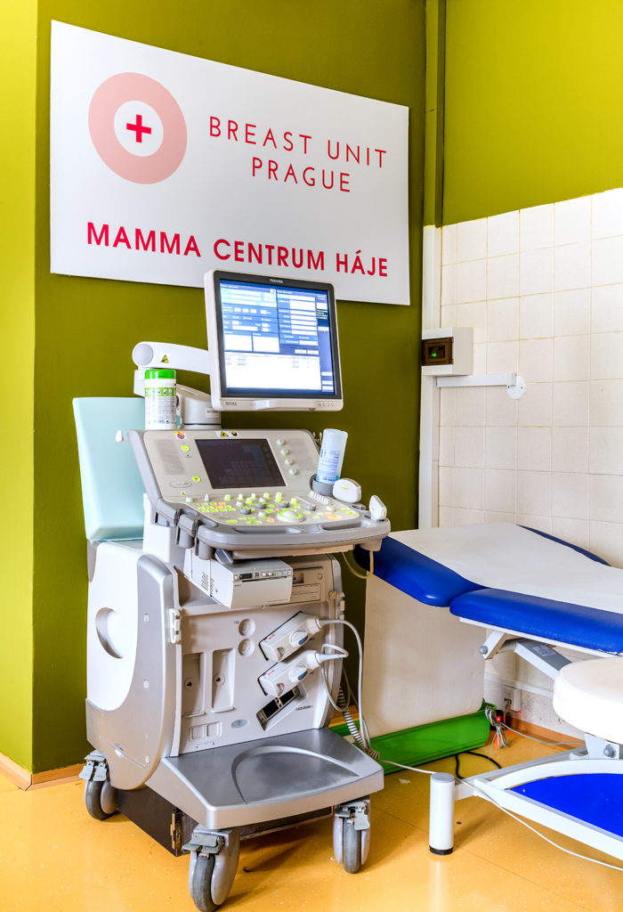

Ultrasound
Ultrasound examination of the breast
Ultrasound examination is basic examination method for women up to 40 years age or a complementary examination to mammography.
Using a dialing system, you will be invited into a booth where you will lock the door behind you, you take it off from the waist up and wait to be asked to enter the office.
Ultrasound Scan it is not painfulcomfortable with him lying on your back, you have hands behind the head ...and turn on your side as directed by the examining physician.
The examination is carried out using ultrasonic probeswhich the doctor swipes across your breasts and watches the image of the breast tissue on the screen. The probe generates ultrasound waves that penetrate the breast tissue and bounce back in different ways, which are picked up by the probe. During the examination, it is necessary to mention any clinical problems that are currently bothering you (palpable lump, secretion, etc.).
The doctor evaluates the findings straight away, dictates the report to the assistant, which you receive immediately after the examination. In some cases it is necessary to perform histological verification by biopsy or puncture - we will make an appointment for these procedures if necessary, the dates are usually within a week.
There is no special preparation before the ultrasound examination, however, it is advisable to avoid the use of body creams and solid antiperspirants. Ultrasound examination can be performed at all stages of the menstrual cycle, preventive check-up it's better to plan after the end of menstruation due to hormonal calming of the breasts and for your comfort during the examination.
Ultrasound is not an X-ray examination, it can therefore be performed in case of difficulties (lump, etc.) also in pregnant and breastfeeding women. However, if you want to have the examination for preventive reasons, it is recommended to have it outside pregnancy and 6 months after the end of breastfeeding.
Ultrasound is ideal complementary examination to mammographybecause a snapshot may not always provide all the information. It helps with so-called dense breasts, where it is not possible to decide what is hidden in the opaque gland. It is also used to resolution of unclear bearing. On the other hand, mammography is fully sufficient for the examination of empty breasts and microcalcifications and ultrasound does not provide any additional useful information.
Only young women can have an ultrasound examination without a previous mammogram. An indispensable role plays ultrasound in the control of cancer response to chemotherapy, examination of axillary nodules and scars after mastectomy and after reconstructive procedures. Most interventional procedures are also performed under ultrasound guidance.
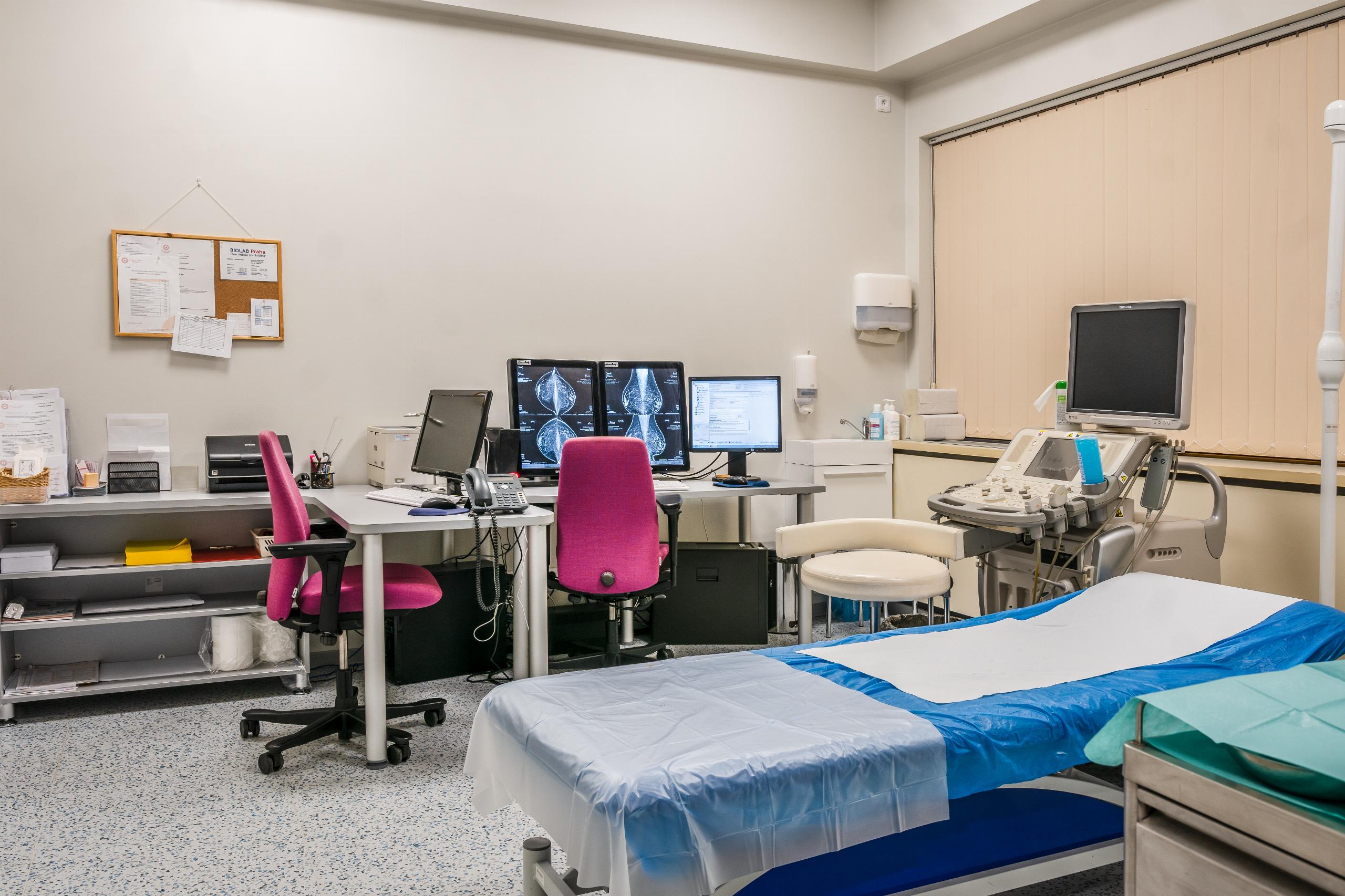
Doctor's office n. 2