Biopsy
The term biopsy means taking a sample from the suspected breast gland to determine the exact histological origin of these changes.
If the doctor decides to biopsy there is no reason for unnecessary concern or panic. This procedure is performed when the nature of the finding cannot be clearly determined from the available imaging methods. We must remember that neither eye an experienced doctor is not a microscope, and therefore a histological analysis of the samples is always necessary for a definitive diagnosis.
We perform three types of biopsies at our centre and the doctor always determines which is best for the individual patient according to the nature of the finding.
These procedures are performed weekly, and the patient is always scheduled for the nearest possible date.
The aim is to determine the exact histological composition of the ambiguous formation in the breast. Biopsy indicated only by a doctor based on unclear results of MG and ultrasound. The patient is booked for biopsy by the radiology assistant directly in the examination room.

Ultrasound
Core-cut biopsy
Core-cut biopsy is one of the most common types of biopsy. We approach it when we have found a circumscribed lesion using imaging methods, for which we cannot determine the exact origin.
Before core-cut biopsy no special preparation required patient, but it is always necessary to check whether the patient is taking lblood thinners and whether he has allergy to local anaesthetics. Even in these cases, biopsy is not contraindicated, only as needed. modifies the diagnostic procedure.
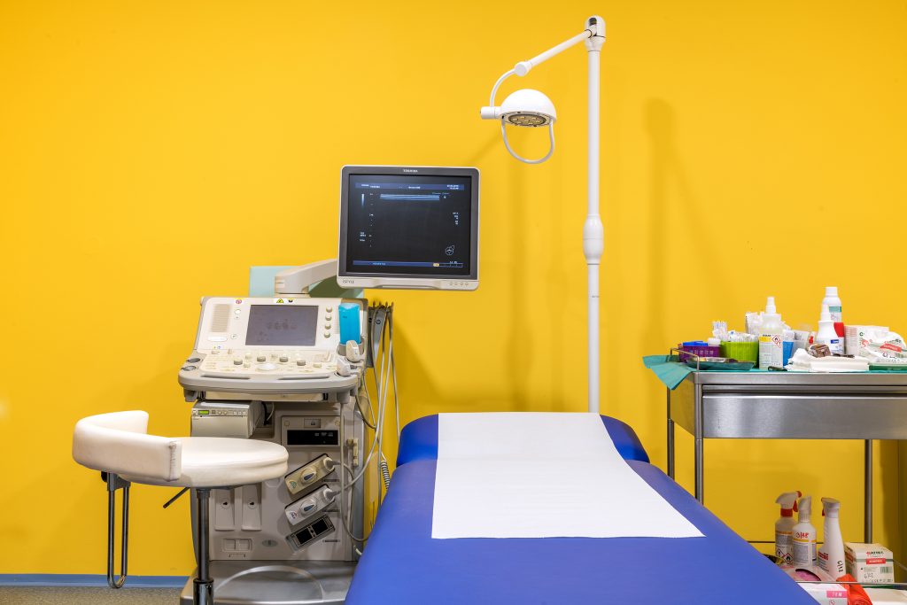
Core-cut biopsy is performed for ultrasound checks, the patient lies comfortably on her back or on her side and the doctor takes the sample.
The power starts by numbing injection site using a local anaesthetic, a thin needle is used to inject a numbing agent into the skin and subcutaneous tissue. This injection may be somewhat uncomfortable, but thanks to the anaesthetic, the patient will not feel any further pain. This is followed by the actual sampling, where the doctor takes with a special biopsy needle, and for ultrasound checks takes samples from the deposit. Now the patient may feel light pressure, but not sharp pain, or an anaesthetic may be added after discussion with the doctor. The biopsy needle will take individual samples, you always need to have several of themso that the laboratory has enough material to make an accurate diagnosis.
That's the end of the performance, now cover the puncture site with sterile squares and squeeze to prevent possible bleeding. After sufficient compression, the puncture site is taped over with a plaster and the patient can go home.
Samples are sent to the laboratory and the result is usually received within 10-14 days. This time of waiting and uncertainty is certainly unpleasant for the patient, the procedure unfortunately cannot be accelerated. As soon as we get the results, we contact the patient by phone and we'll agree on a course of action.
Vacuum biopsy under ultrasound
Vacuum biopsy under ultrasound is performed in case of, if the bearing is not completely unambiguous, or if it is rather a precinct with not very clear contours.
The procedure is not fundamentally different from core-cut biopsyis performed lying down in the ultrasound room. The site is numbed with a local anaesthetic and the doctor uses a needle to take the sample. After the needle is inserted and the first samples are taken, the needle is only rotated, so we can obtain samples from multiple sites from a single puncture.
After the collection is completed, the injection site is again compressed, then covered with adhesive stitches and a sterile patch.
Stereotactic vacuum biopsy
Stereotactic vacuum biopsy is performed in cases where it is necessary to verify the so-called microcalcification (see glossary). Since microcalcifications are not visible on ultrasound, this performance for mammography checks. Successful removal of microcalcifications depends on their correct targeting, so first the radiology assistant sits the patient at the mammography machine and locates the site from which the removal will be performed. The breast is then compressed as in a routine mammogram and a control image is taken to verify that we've found the correct location. Now it's important that the patient She wasn't moving and remained seated in the same position throughout the performance.
Instead of the doctor again numb with a local anaesthetic and then the needle is inserted. The removal itself is also not painfulby rotating the needle, the doctor achieves the collection of several tissue samples with the desired microcalcifications. In some cases, the site is marked with a clip for easier identification of the site in the future.
The injection site is compressed to prevent possible bleeding and then treated with adhesive stitches and a sterile patch.
Biopsy FAQs
-
Will the biopsy hurt?
The procedure itself is not painful, it is performed after the site of removal is numbed. The actual application of anaesthetic with a thin needle into the skin and subcutaneous tissue can be uncomfortable and may sting a little.
-
How long will the biopsy take?
The whole examination and collection takes approximately 20 minutes, the collection itself takes about a minute.
-
What are the performance risks?
We must always check whether the patient is allergic to the anaesthetic, disinfectant or patch to prevent an allergic reaction.
After the procedure, a minor bruise usually occurs, but in rare cases, when a larger vessel is disrupted during the procedure, a more significant hematoma may occur.
The risk of introducing infection into the breast is negligible (max. 0.05%)
-
Can I go to work after the biopsy?
After the biopsy, we recommend a relatively restful regime, but this does not mean strictly lying in bed. But definitely avoid heavy physical work (lifting heavy loads, washing windows, playing sports). If you do physically demanding work, you can go to work after exercising.
-
Can I bathe immediately after the procedure?
You can shower on the day of the procedure, bathe in the bath, swimming pool, etc., we recommend 3-5 days, when the injection site will be calm and the stroma will form.
-
Will the injection site hurt after the anesthetic wears off?
It may happen that a bruise forms on the skin after removal, so you may feel pain as if you have hit the spot. We recommend that you ice the site, or take a regular anaesthetic that you take for pain (not Acylpyrine etc., which causes blood thinning and could therefore cause the puncture site to bleed again).
-
How long before I know the results?
We will receive the results from the laboratory usually in 10-14 days and we will contact the patient by phone.
-
What is the procedure after the results come in?
If the result of the biopsy requires a solution, we will arrange subsequent treatment and surgery. If the patient wants to arrange the treatment herself, we will issue the necessary documentation.
If the biopsy result does not require any solution, we will arrange a follow-up with the patient.

